Quick answer
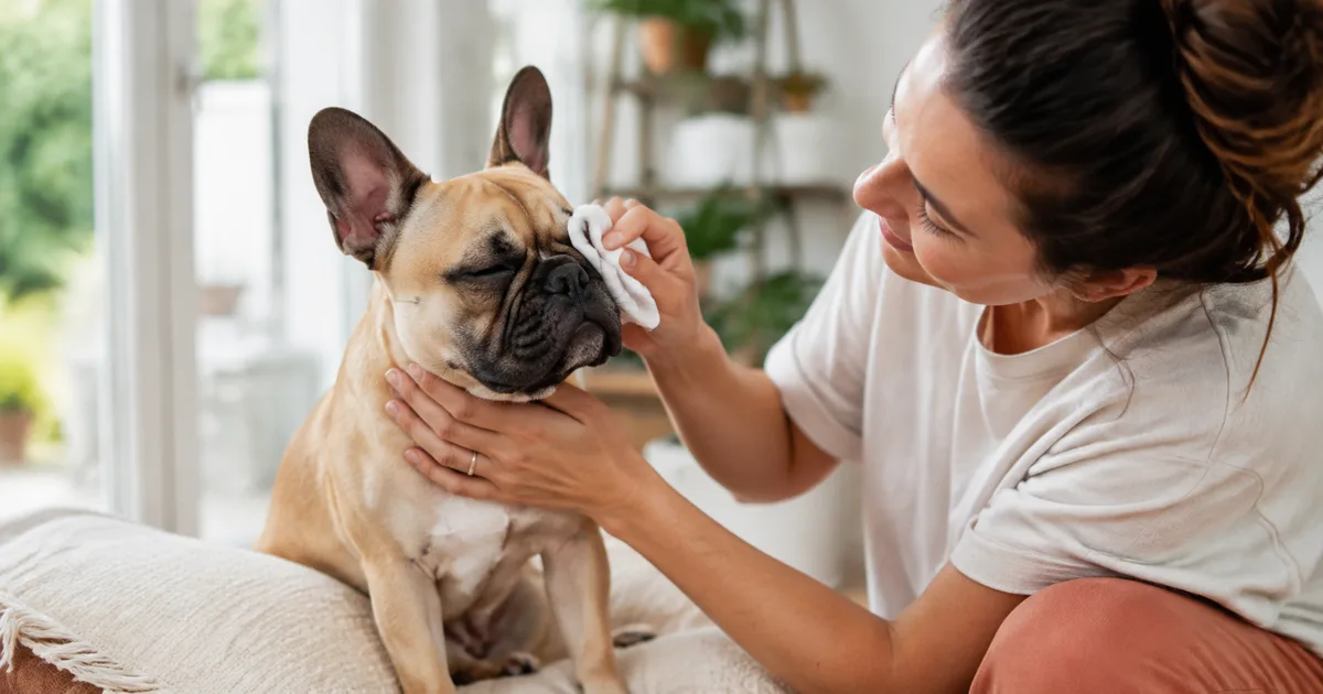
A small amount of dried material at the inner corner of a comfortable eye can sometimes be cleaned gently from the surrounding fur. Use clean gauze or a soft pad moistened with sterile saline eye wash, or follow a cleaning method your veterinarian has approved. Soften crusts before wiping, use a fresh pad for each eye, and keep the skin folds dry afterward.
Cleaning is not a treatment for the cause of discharge. French Bulldogs have prominent, relatively exposed eyes and facial anatomy that can reduce corneal protection. New, one-sided, persistent, thick, foul-smelling, or increasing discharge deserves veterinary advice, even if your Frenchie is not obviously painful.
Seek same-day veterinary care for squinting, holding an eye closed, marked redness, rubbing, swelling, a cloudy or blue-white cornea, a visible spot or wound, sudden vision difficulty, blood, trauma, or an eye that looks larger, smaller, or more prominent than usual. Eye disease can progress quickly, and some flat-faced dogs show less pain than expected despite serious corneal injury.
When eye discharge is urgent
| What you notice | What to do |
|---|---|
| Eye displaced or bulging after trauma, deep cut, penetrating object, or active bleeding | Emergency veterinary care now |
| Closed eye, repeated squinting, severe rubbing, cloudy or blue-white cornea, visible crater or spot, sudden vision change | Same-day urgent eye examination |
| Redness with thick discharge, eyelid swelling, increasing pain, or rapid worsening | Contact a veterinarian promptly, usually the same day |
| Persistent discharge from one eye, recurrent tear staining with odor or sore skin, or discharge that keeps returning | Arrange a veterinary appointment |
| Small amount of dried material with a fully open, white, comfortable eye and no recurrence | Gently clean the surrounding fur and continue observing |
Do not wait for the discharge to change color before seeking care. A painful eye or abnormal cornea is more important than whether the material looks clear, yellow, green, or brown.
Why French Bulldog eyes need extra attention
French Bulldogs are brachycephalic, meaning their skulls are shortened. This facial shape can leave the eyes more prominent and the sockets relatively shallow. Some dogs do not close the eyelids completely with every blink, have reduced corneal sensitivity, or develop hair contact from nasal folds or inward-rolling eyelids. These features can increase exposure, dryness, irritation, and injury.
The American College of Veterinary Ophthalmologists describes this group of problems as brachycephalic ocular syndrome. Affected dogs may have excessive tearing, redness, discharge, poor blinking, corneal ulcers, pigment, or chronic surface disease. Reduced corneal sensitivity matters because a Frenchie may not squint dramatically even when the cornea is injured.
A study of 93 brachycephalic dogs referred for ophthalmic examination found corneal ulcers, pigmentation, fibrosis, and entropion among the frequent abnormalities. This was a referral population and does not predict the risk for every French Bulldog, but it shows why persistent discharge should not automatically be treated as a harmless breed trait.
What different discharge patterns can tell you
The appearance provides context, but color and texture cannot diagnose the cause.
Clear and watery
Clear overflow may occur with irritation, wind, dust, hair touching the eye, an eyelid problem, a blocked tear-drainage pathway, pain elsewhere in the eye, or excessive tear production. Sudden watering from one eye raises more concern for a foreign object, scratch, or corneal ulcer than long-standing mild staining under both eyes.
White, gray, or stringy mucus
Mucus can occur when the tear film is unstable or the eye surface is irritated. Dry eye, known as keratoconjunctivitis sicca (KCS), may produce sticky or stringy material because the watery component of tears is reduced. Dry eye can also cause redness, corneal inflammation, ulcers, scarring, and pigment.
Yellow or green discharge
Thick yellow or green material reflects inflammatory cells and debris, but it does not prove that a simple bacterial infection is the primary problem. Dry eye, an ulcer, an eyelid abnormality, a foreign body, or another condition may allow secondary infection or produce similar discharge. Antibiotic drops selected without an examination may fail to address the cause.
Reddish-brown staining
Tears left on light-colored fur can create reddish-brown staining. The stain itself may be cosmetic, but constant wetness can irritate the skin and create odor. New or heavy staining may reflect excessive tearing, poor drainage, eyelid or hair contact, or eye discomfort. Cleaning the fur does not determine why tears are overflowing.
Blood-tinged discharge
Blood may come from trauma, severe inflammation, a mass, a foreign object, or bleeding around the eye. It warrants prompt veterinary assessment, especially when accompanied by pain, swelling, cloudiness, or a change in vision.
One eye versus both eyes
Discharge from only one eye makes a local problem more likely, such as a foreign object, corneal injury, blocked tear duct, abnormal eyelash, eyelid problem, or focal infection. Bilateral discharge may occur with dry eye, environmental irritation, allergy-associated inflammation, or facial anatomy affecting both sides.
This distinction is helpful for the history, but it is not absolute. Dry eye can be worse on one side, and trauma or an ulcer can eventually cause broader inflammation. Record which eye changed first and whether the signs started suddenly.
Common causes in French Bulldogs
Corneal irritation or ulcer
The cornea is the clear surface at the front of the eye. A scratch, foreign material, hair contact, reduced tear protection, chemical exposure, or infection can damage it. Signs may include tearing, discharge, redness, squinting, rubbing, light sensitivity, or a cloudy area. Brachycephalic dogs are particularly vulnerable to serious corneal disease.
A corneal ulcer cannot be confirmed from a photograph or discharge color. Veterinarians commonly use fluorescein dye, which adheres to damaged corneal tissue, to identify an ulcer and assess its extent. Deep or infected ulcers can threaten vision and may require intensive medical care or surgery.
Dry eye
Dry eye occurs when the tear film does not adequately protect the ocular surface. Cornell University notes that canine KCS can cause redness, irritation, thick green discharge, and a risk of painful corneal ulcers. Chronic disease can lead to blood-vessel growth, scarring, pigmentation, and vision loss.
Treatment usually involves long-term prescription medication that supports tear production or replaces part of the tear film, along with management of inflammation or infection when present. Frequent cleaning alone cannot restore the tear film.
Entropion, eyelashes, or facial hair contact
Entropion is an inward roll of the eyelid that allows skin or hair to rub the cornea. Misplaced eyelashes and hair from a prominent nasal fold can also irritate the eye. The result may be tearing, mucus, redness, ulcers, or progressive corneal pigment. Some flat-faced dogs show surprisingly little discomfort.
Read our French Bulldog entropion guide for how veterinarians assess eyelid position and choose temporary or surgical treatment.
Conjunctivitis
Conjunctivitis means inflammation of the membrane lining the eyelids and covering the white of the eye. It can occur with allergy, irritation, dry eye, eyelid disease, infection, trauma, or disease elsewhere in the eye. Redness and discharge describe inflammation; they do not identify its cause.
Because treatments differ, “pink eye” should not be treated with a human product or an old veterinary prescription. The veterinarian may need to rule out an ulcer, dry eye, or abnormal eye pressure before selecting medication.
Tear-drainage problems
Tears normally move through small openings near the inner eyelids and into the nasolacrimal drainage system. Narrowing, inflammation, debris, or facial anatomy can reduce drainage and cause overflow onto the face. A veterinarian may use fluorescein dye or flush the system when a drainage problem is suspected.
Cherry eye and third-eyelid disease
Cherry eye is prolapse of the tear-producing gland of the third eyelid. It appears as a smooth pink or red swelling near the inner corner and may be associated with irritation and discharge. Because the gland contributes to tear production, treatment usually aims to preserve and reposition it rather than remove it. See our French Bulldog cherry eye guide for examination and treatment details.
Skin-fold irritation
Constant moisture beneath the eyes can irritate facial skin and folds, creating redness, odor, or surface infection. The eye and the skin may both need assessment. Keeping the fold clean and dry can support comfort, but an ongoing wet track should prompt investigation of the eye and tear drainage. Our French Bulldog skin-fold guide covers safe fold care and warning signs.
How to clean around the eyes safely
Only clean at home when the eye is open, appears comfortable, and has no trauma, marked redness, cloudiness, or visible injury.
- Wash and dry your hands. Prepare clean gauze or soft lint-free pads. Avoid a fluffy material that leaves fibers behind.
- Moisten the pad. Sterile saline eye wash is a useful option for cleaning around the eye. A product prescribed or approved by your veterinarian should be used as directed.
- Soften dried material. Hold the damp pad gently against the crust on the fur for a short time. Do not press on the eyeball or scrape hardened debris.
- Wipe away from the eye. Use a gentle outward motion across the surrounding fur. Do not rub back and forth across the eyelid margin.
- Use a new pad for each eye. This reduces transfer of discharge and debris from one side to the other.
- Dry the skin and folds. Pat the surrounding fur dry without rubbing. Persistent dampness can irritate the skin.
- Stop if your Frenchie resists or becomes painful. A closed or painful eye needs an examination rather than more cleaning.
If your veterinarian has prescribed drops, clean visible discharge first unless the clinic instructs otherwise. Keep the applicator tip from touching the eye, eyelids, fur, hands, or another surface.
Products and home remedies to avoid
Do not place the following in or near the eye unless a veterinarian who examined your dog specifically directs it:
- human redness-relief or allergy eye drops;
- leftover antibiotic, anesthetic, or steroid drops;
- contact-lens cleaning or disinfecting solution;
- hydrogen peroxide, alcohol, vinegar, or household cleaners;
- essential oils, herbal tea, or homemade mixtures;
- tear-stain bleach, powders, or abrasive wipes close to the eye;
- ointments or skin creams that are not labeled for ophthalmic use.
Old steroid eye medication is especially risky. Merck Veterinary Manual states that corticosteroids are contraindicated when a corneal ulcer is present because they can interfere with healing and worsen infection. The only safe way to know whether an ulcer is present is through an eye examination and appropriate testing.
What if shampoo or a chemical enters the eye?
Begin flushing immediately with sterile saline or clean lukewarm water. For a corrosive exposure, Merck recommends prolonged irrigation and subsequent veterinary examination with fluorescein staining to check the cornea. Contact a veterinarian or animal poison service while flushing or immediately afterward and take the product packaging with you.
Do not try to neutralize one chemical with another. Do not delay irrigation while searching for a preferred eye product.
What the veterinarian may test
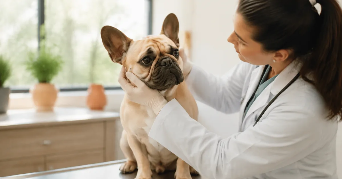
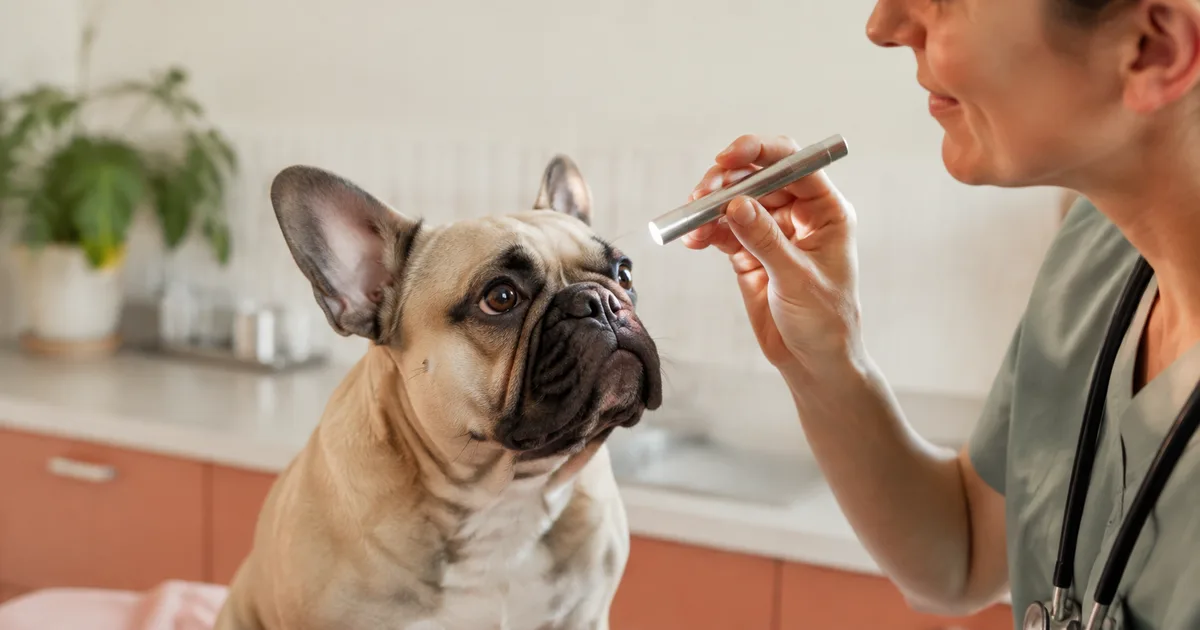
An eye examination considers the eyelids, lashes, facial folds, third eyelid, conjunctiva, cornea, pupils, lens, and structures inside the eye. Common tests include:
- Schirmer tear test: measures the watery component of tear production.
- Fluorescein stain: checks for corneal ulcers and may help assess tear drainage.
- Tonometry: measures pressure inside the eye to investigate glaucoma or uveitis.
- Magnified examination: helps identify abnormal hairs, small foreign material, ulcer depth, and corneal changes.
- Cytology or culture: may be used for selected severe, infected, unusual, or non-healing conditions.
- Nasolacrimal evaluation: assesses whether tears can drain through the normal pathway.
Testing order matters. Tear production and corneal staining are often checked before drops or extensive cleaning change the eye surface.
Treatment depends on the cause
An ulcer may need protective medication, antimicrobial treatment, pain control, repeated checks, and sometimes surgery. Dry eye often requires long-term tear-stimulating or lubricating medication. Entropion or persistent hair contact may require eyelid correction. A blocked tear pathway may need flushing or treatment of local inflammation. Glaucoma and uveitis require prompt, cause-specific care.
This is why cleaning and “natural” drops cannot serve as universal eye-discharge treatment. They may remove material from the fur while the underlying condition continues.
Frequently asked questions
Is a little eye discharge normal for a French Bulldog?
A small amount of dried material after sleep can occur in an otherwise comfortable eye. Recurrent wetness, increasing material, odor, red skin, one-sided discharge, redness, cloudiness, or squinting is a reason to contact your veterinarian.
Does green discharge always mean infection?
No. Green or yellow material can appear with dry eye, corneal disease, eyelid problems, and secondary infection. An examination determines whether antimicrobial treatment is appropriate and whether another condition is driving the discharge.
Can I use human saline solution?
Use a sterile product intended as simple eye wash, ideally one your veterinarian recommends. Contact-lens disinfecting solutions and medicated human drops are different products and should not be substituted.
Why does my Frenchie have discharge from only one eye?
One-sided discharge may occur with a foreign object, scratch or ulcer, abnormal hair, blocked tear drainage, eyelid disease, or another local problem. Persistent unilateral discharge should be examined.
What to remember
Gentle cleaning can keep the fur and folds comfortable, but it cannot diagnose or treat the reason eye discharge is present. French Bulldogs can have significant corneal disease with subtle pain behavior, so prioritize the condition of the eye over the color of the discharge. Seek prompt care for squinting, redness, rubbing, cloudiness, injury, swelling, blood, or vision change.
Explore the rest of our French Bulldog eye-health guides for related eyelid and third-eyelid conditions.
Sources
- American College of Veterinary Ophthalmologists: Understanding brachycephalic ocular syndrome
- American College of Veterinary Ophthalmologists: How to clean your pet's eyes
- Cornell University College of Veterinary Medicine: Keratoconjunctivitis sicca in dogs
- Merck Veterinary Manual: Disorders of the conjunctiva in dogs
- Merck Veterinary Manual: Physical examination of the eye in animals
- Merck Veterinary Manual: Anti-inflammatory agents in animals
- Costa et al.: Clinical signs of brachycephalic ocular syndrome in 93 dogs
- Spornberger et al.: Tear-film characteristics in French Bulldogs and non-brachycephalic dogs
This article is for education and triage. It cannot diagnose an eye condition or replace veterinary examination and treatment.