Quick answer
Entropion means part of the eyelid rolls inward, allowing eyelid hair or nearby facial hair to rub the clear surface of the eye (the cornea). That constant friction can cause tearing, squinting, ulcers, pigment, scarring, and—in severe untreated cases—loss of vision.
For a French Bulldog with a painful, closed, cloudy, or suddenly changed eye, contact a veterinarian the same day. Eye problems can look alike from the outside, and a corneal ulcer cannot be ruled out at home. Lubricants may protect the surface temporarily when prescribed, but they do not correct an eyelid that keeps rolling inward. Established entropion commonly requires surgery; selected puppies may first receive temporary eyelid-tacking sutures while their face matures.
When is entropion an emergency?
Use this table for triage, not diagnosis:
| What you see | What to do |
|---|---|
| Eye held shut, marked squinting, repeated pawing, or sudden light sensitivity | Arrange a same-day veterinary examination |
| Blue, white, or cloudy spot on the cornea; blood; a visible wound; sudden bulging; or an object in the eye | Seek urgent or emergency veterinary care now |
| New thick yellow-green discharge with redness or pain | Contact a veterinarian the same day |
| Mild tearing that persists or keeps returning, even if your Frenchie acts comfortable | Book a prompt examination; brachycephalic dogs may show less obvious discomfort |
| Mild swelling after eyelid surgery that is stable or improving | Follow the surgeon’s discharge plan and scheduled recheck |
| Increasing swelling, bleeding, incision discharge, worsening squinting, cloudiness, or broken sutures after surgery | Call the surgical clinic promptly |
Do not put redness-relief drops, leftover antibiotics, or any steroid-containing eye medication into a painful eye unless the examining veterinarian specifically prescribed it for this episode. Some products can delay appropriate care, and topical corticosteroids are contraindicated when a corneal ulcer is present.
What entropion does to a Frenchie’s eye
A normal eyelid margin sits against the eye and spreads tears with each blink. With entropion, the margin turns inward. The resulting contact may be intermittent at first—perhaps worse when a dog is tired or squinting—or continuous.
This is more than a cosmetic wrinkle. Repeated hair-to-cornea contact acts like ongoing abrasion. The cornea may become inflamed or ulcerated; chronic cases can develop blood vessels, dark pigment, or scar tissue that interferes with vision. The American College of Veterinary Ophthalmologists (ACVO) notes that untreated entropion can cause ulcers, perforation, scarring, and vision deficits.
French Bulldogs also have brachycephalic—or short-faced—eye anatomy. Their prominent eyes, relatively exposed corneas, facial folds, incomplete blinking in some dogs, and tear-film problems can reduce the eye’s margin for error. Entropion is only one part of that picture. In a referral-hospital study of 93 brachycephalic dogs, including 38 French Bulldogs, corneal ulceration, pigmentation, fibrosis, and entropion all appeared among the recorded ocular findings. That study describes a referred clinical population, so its percentages should not be read as the prevalence in every Frenchie.
Why French Bulldogs develop entropion
Entropion is a description of eyelid position, not one single disease. A veterinarian needs to identify what is pulling or pushing the lid inward before choosing treatment.
Developmental or conformational entropion
The shape of the skull, eyelid opening, surrounding skin, and facial folds can make the eyelid sit incorrectly. This primary form may become apparent in a puppy or young adult and can affect one or both eyes. In short-faced dogs, the inner—or medial—part of the eyelid deserves particular attention because nearby nasal-fold hair may also contact the cornea.
Spastic entropion caused by eye pain
Pain makes a dog squeeze the eyelids tightly. That spasm can roll the lid inward, creating a damaging loop: an ulcer or other painful condition causes squinting; squinting worsens the inward roll; the inward roll causes more corneal rubbing.
This distinction matters. The Merck Veterinary Manual advises managing spastic entropion temporarily while treating the underlying keratitis or ulcer. Once the pain resolves, permanent eyelid surgery may no longer be necessary—or the examination may reveal a smaller underlying conformational problem.
Acquired entropion
Less commonly, eyelid scarring, trauma, inflammation, loss of support around the eye, or another structural change alters lid position later in life. Treating the cause and planning surgery can be more complex in these cases.
Signs owners may notice
Entropion is not always easy to see beneath a Frenchie’s folds. Watch for:
- Squinting, frequent blinking, or holding one eye partly or fully closed.
- Tearing or a wet track below the eye.
- Clear, mucus-like, or colored discharge.
- Pawing at the face or rubbing on furniture or carpet.
- Redness around the eye.
- Sensitivity to light or reluctance to have the head touched.
- A blue-white, dull, hazy, or irregular area on the cornea.
- New brown or black pigment spreading across the clear cornea.
- An eyelid edge that disappears inward, or hair visibly touching the eye.
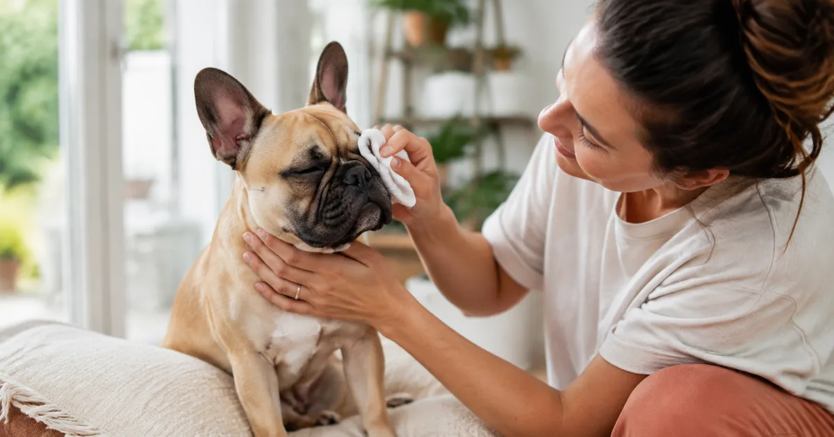
Some brachycephalic dogs show little obvious pain, so a cheerful Frenchie can still have corneal disease. Persistent discharge is a symptom, not a diagnosis. Our French Bulldog eye-discharge guide explains what can be cleaned safely while you arrange care.
How a veterinarian diagnoses it
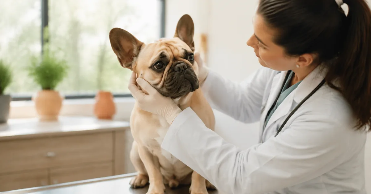
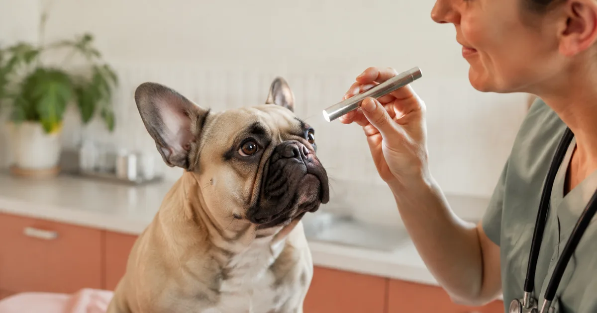
Diagnosis begins with observing your dog before the eyelids are handled. A veterinarian evaluates facial symmetry, the shape of the eyelid opening, blink coverage, facial folds, hair contact, and whether the lid position changes when your Frenchie relaxes.
A complete eye assessment may include:
- Fluorescein staining: dye adheres to exposed corneal tissue and helps identify an ulcer.
- Schirmer tear testing: a paper strip measures the watery portion of tear production.
- Tonometry: an instrument measures pressure inside the eye when appropriate.
- Magnified examination: the cornea, eyelid margin, lashes, conjunctiva, and third eyelid are inspected for other causes of irritation.
These tests help separate entropion from dry eye, abnormal eyelashes, facial-fold hair contact, infection, trauma, glaucoma, and other painful conditions. Cherry eye is different: it is prolapse of the third-eyelid gland, seen as a pink or red mass at the inner corner. Read our French Bulldog cherry-eye guide for that condition.
If pain-induced spasm makes the lid position difficult to judge, the veterinarian may first protect the cornea, control pain, or temporarily tack the lid outward, then reassess its natural position. Complicated anatomy, a deep or nonhealing ulcer, previous eyelid surgery, or uncertain surgical planning may justify referral to a board-certified veterinary ophthalmologist.
Treatment options: what each one can and cannot do
| Approach | When it may be used | Important limitation |
|---|---|---|
| Veterinary-prescribed lubricant and treatment for corneal injury | Protects the surface and treats associated disease while a plan is made | Does not permanently reposition an inward-rolling lid |
| Temporary eyelid tacking | Often considered for a growing puppy or spastic entropion | Sutures are temporary; reassessment is essential and permanent correction may still be needed |
| Injectable filler | A specialist may consider it for selected patients that are poor surgical candidates | Effect is temporary and suitability depends on anatomy and clinician experience |
| Corrective eyelid surgery | Established conformational entropion or ongoing corneal contact | Requires anesthesia, precise planning, aftercare, and sometimes revision |
Temporary tacking in puppies
Small sutures roll the eyelid away from the cornea while the puppy grows or while ocular pain settles. ACVO explains that young animals may be managed this way to avoid removing tissue before skull development is complete. Tacking protects the eye, but it is not permission to wait without follow-up: sutures can loosen, irritation can recur, and some dogs still need definitive surgery after maturation.
Definitive surgery
The common principle is to remove or reshape a carefully measured strip of skin near the affected margin so the lid sits in a healthier position. A frequently used technique is the modified Hotz-Celsus procedure, but there is no universal operation for every Frenchie. The surgeon may need to address the inner corner of the eyelids, an oversized eyelid opening, nasal-fold hair, abnormal lashes, or a corneal lesion as part of an individualized plan.
Precision matters: too little correction leaves hair rubbing the eye; too much can turn the lid outward, prevent full closure, or expose the cornea. A staged or conservative correction may therefore be safer. Ask how frequently the surgeon operates on brachycephalic eyelids and whether ophthalmology referral would improve planning.
What recovery usually looks like
Recovery varies with the procedure, the condition of the cornea, and your dog’s health. The surgeon’s written instructions override any general timeline.
| Time | What may be expected | Your priorities |
|---|---|---|
| Surgery day | Sleepiness after anesthesia; mild incision redness or swelling | Quiet supervision, prescribed medication, and no access to the incision |
| First 24–72 hours | Bruising or swelling can be most noticeable; comfort should be stable or improving | Keep the cone on, restrict activity, and check both eye and incision several times daily |
| Days 3–7 | Swelling should trend down; the lid may still look uneven while tissues heal | Continue medications and short leash walks only; call if pain or discharge increases |
| About days 7–14 | Many dogs have a planned recheck; external sutures may be removed if healing allows | Do not remove sutures or stop the cone without the clinic’s approval |
| Following weeks | The lid position becomes easier to judge as swelling resolves | Attend every recheck and monitor for renewed rubbing, tearing, or inward rolling |
A cosmetic verdict in the first few swollen days is premature. The immediate goal is a protected, comfortable cornea—not perfectly symmetrical eyelids in a photograph.
Aftercare at home
- Keep the Elizabethan collar on continuously. Scratching can damage the cornea or pull out sutures. Inflatable collars may not extend far enough to protect a Frenchie’s eye; use the device approved by the surgeon.
- Give each medication exactly as labeled. Wash your hands, avoid touching the bottle tip to the eye or hair, and leave the interval your clinic recommends between multiple drops or ointments.
- Keep the incision dry. Do not bathe your dog, apply skin products, use peroxide, or place a compress over the area unless instructed.
- Restrict activity. Use a leash for toilet breaks, prevent rough play and jumping, and separate pets that might lick or paw the face.
- Check progress in consistent light. A daily photo can reveal a trend, but it does not replace the scheduled examination.
- Protect breathing as well as the eye. French Bulldogs are brachycephalic. Follow the clinic’s anesthesia-discharge instructions closely, keep your dog cool, and urgently report difficult or noisy breathing that is worse than their normal baseline.
Never restart old eye medicine on your own. A drug prescribed for a previous episode may be wrong for a new ulcer, and a bottle tip can become contaminated.
Possible complications and long-term outlook
Most appropriately treated dogs are more comfortable once hair no longer rubs the cornea. Prognosis depends partly on how much damage existed before correction: fresh irritation may resolve, whereas established pigment or scar tissue may not disappear completely.
Potential complications include infection, bleeding, suture irritation, wound opening, residual entropion, overcorrection, incomplete eyelid closure, and recurrence. A revision does not necessarily mean the first operation was careless; cautious correction can be deliberate when excessive tissue removal would threaten closure.
Continue checking both eyes after recovery. Conformation can affect more than one portion of the eyelids, and tear-film or corneal problems can coexist even after the mechanical rubbing is fixed. Routine fold care may reduce nearby skin irritation, but cleaning facial folds cannot cure entropion.
Questions to ask before surgery
- Is this primary conformational entropion, pain-related spastic entropion, or both?
- Is the cornea ulcerated or scarred, and does that change the timing or prognosis?
- Which part of the eyelid is affected, and are nasal folds or abnormal lashes also contacting the eye?
- Is temporary tacking reasonable, or is definitive correction recommended now?
- Which procedure is planned, and might more than one eyelid procedure be needed?
- What medications, cone type, activity limits, and recheck dates should I plan for?
- Which changes require an emergency visit rather than waiting for the recheck?
- Would referral to a board-certified veterinary ophthalmologist help this case?
You can locate a specialist through the ACVO ophthalmologist directory.
Frequently asked questions
Can a puppy outgrow entropion?
Some young dogs improve as their facial structures develop, which is why temporary tacking may be chosen. It is not safe to simply “wait and see” while hair rubs the cornea. The eye still needs protection and scheduled reassessment.
Can drops cure entropion?
No drop changes eyelid anatomy. Prescribed lubricants, antibiotics, pain relief, or other medication may protect and treat the eye before or after surgery, but established mechanical rolling usually needs a mechanical correction.
Does entropion always affect both eyes?
No. It can involve one or both eyes and only part of an upper or lower lid. The veterinarian should examine both eyes because subtle contact may be present on the side that looks normal at home.
How much does entropion surgery cost?
There is no responsible universal figure. Cost varies with location, surgeon, testing, one versus both eyes, complexity, corneal treatment, medications, and rechecks. Ask for a written estimate that separates eyelid correction from treatment of any ulcer.
Can entropion return after surgery?
Yes, although many dogs do well after one definitive correction. Recurrence or residual rolling can occur as swelling resolves, a puppy grows, scar tissue changes, or another section of the lid becomes involved. Keep every recheck so the final position is judged after healing—not just immediately after surgery.
Bottom line
French Bulldog entropion is a mechanical source of corneal injury, not merely a watery-eye problem. Squinting, a closed eye, cloudiness, or repeated rubbing deserves same-day veterinary attention. Careful examination separates conformational entropion from pain-induced eyelid spasm and checks for an ulcer. Temporary tacking can protect selected puppies or painful eyes, while established entropion often needs precisely planned surgery. Consistent cone use, medication, activity restriction, and follow-up are the foundation of a safe recovery.
Sources
- American College of Veterinary Ophthalmologists: Entropion
- American College of Veterinary Ophthalmologists: Brachycephalic ocular syndrome
- Merck Veterinary Manual: Eyelids in animals
- Merck Veterinary Manual: Physical examination of the eye in animals
- Merck Veterinary Manual: Disorders of the eyelids in dogs
- Costa et al., Clinical signs of brachycephalic ocular syndrome in 93 dogs
- Spornberger et al., Tear film characteristics in French Bulldogs
- VCA Animal Hospitals: Post-operative instructions in dogs
This article is educational and does not diagnose your dog or replace veterinary care. Eye pain and corneal changes can be time-sensitive; contact a veterinarian for advice based on an examination.